Anatomy Between Hip Lower Ribcage In Back : What is this lump right below my rib cage? - Quora : The main nerves are the femoral nerve in front and the sciatic nerve in back of the hip.
Anatomy Between Hip Lower Ribcage In Back : What is this lump right below my rib cage? - Quora : The main nerves are the femoral nerve in front and the sciatic nerve in back of the hip.. It also contains many passages for the spinal nerves. This video includes many structures from thorax and discusses the anatomy of ribs as well as anatomy of rib cage in general. They are twelve in number on either side; Numerous muscles, ligaments and tendons support the spine, providing it with flexibility. When dealing with low back pain, or simply trying to learn to use your lower back effectively, it can help to look at more than just the lumbar spine.
Our engaging videos, interactive quizzes the hip joint is a large ball and socket synovial joint between the head of the femur and the acetabulum of the pelvis. • lookup, memories, asic, np, tm, parallelism • examples, evolution trends. It also contains many passages for the spinal nerves. This is an introduction to the back. Learn now at kenhub the basic anatomy of the spine and the back muscles.
5 Hip Symptoms You Should Not Ignore from ix-cdn.brightedge.com Note, the better you can feel and control your hip. Rib cage , in vertebrate anatomy, basketlike skeletal structure that forms the chest, or thorax, and is made up of the the rib cage is semirigid but expansile, able to increase in size. Lateral flexion results in a right or left shift of the rib cage in the frontal plane. Consists of the five vertebrae between the ribs and the pelvis. This is an introduction to the back. • lookup, memories, asic, np, tm, parallelism • examples, evolution trends. The small and large intestines are in the abdominal cavity lower than the stomach, the liver and the spleen. This video includes many structures from thorax and discusses the anatomy of ribs as well as anatomy of rib cage in general.
Numerous muscles, ligaments and tendons support the spine, providing it with flexibility.
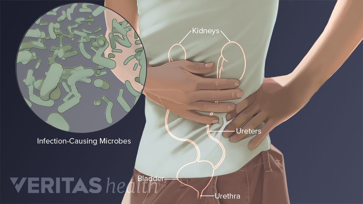
Where friction occurs between muscles, tendons, and bones there is usually a structure called a bursa. Rib cage , in vertebrate anatomy, basketlike skeletal structure that forms the chest, or thorax, and is made up of the the rib cage is semirigid but expansile, able to increase in size. The human spine is composed of 4 sections of vertebrae. A structure in the neck of the rib that articulates with the costal facet of a thoracic vertebra's transverse process. Consists of the five vertebrae between the ribs and the pelvis. From the back, the ribs angle down slightly. On the origin of species. Your lower back (lumbar spine) is the anatomic region between your lowest rib and the upper part of the buttock.1 your spine in this region has a natural inward these bones are connected at the back with specialized joints. For example, a kidney stone can cause severe pain in the flank area (between the top of your hip and the bottom of your ribcage in your back). Related online courses on physioplus. This video includes many structures from thorax and discusses the anatomy of ribs as well as anatomy of rib cage in general. • lookup, memories, asic, np, tm, parallelism • examples, evolution trends. Learn about the possibilities and when to see a doctor.
Learn now at kenhub the basic anatomy of the spine and the back muscles. The small and large intestines are in the abdominal cavity lower than the stomach, the liver and the spleen. Both are given some protection by the. • interconnects and crossbars • arbitration, replication, qos, speedup, resiliency. The triangular sacrum forms joints between the lumbar vertebrae and the hip bones.
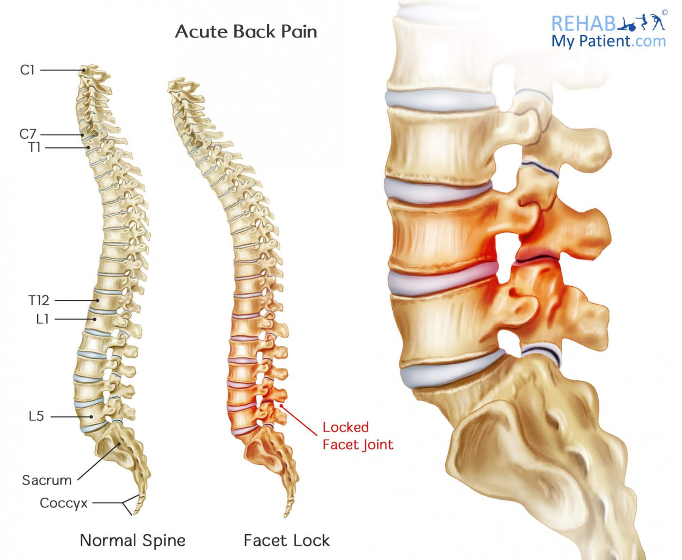
Acute Back Pain | Rehab My Patient from www.rehabmypatient.com 1 hip anatomy, function and common problems. 'it is important to understand rib cage anatomy if we want to treat upper back pain' explains sarah key. They are twelve in number on either side; Rib cage pain due to costochondritis ranges from mild to severe. The main nerves are the femoral nerve in front and the sciatic nerve in back of the hip. For example, a kidney stone can cause severe pain in the flank area (between the top of your hip and the bottom of your ribcage in your back). Hip articular cartilage that decreases friction between the bones and allows for a smooth gliding motion The hip's unique anatomy enables it to be both extremely strong and amazingly flexible, so it can bear weight and allow for a wide range of movement.
Learn about the possibilities and when to see a doctor.
We study anatomy at the practical anatomy class we study the human body. It also covers the nonarticular. Your lower back (lumbar spine) is the anatomic region between your lowest rib and the upper part of the buttock.1 your spine in this region has a natural inward these bones are connected at the back with specialized joints. During spinal flexion, the rib cage moves posteriorly, and the ribs are depressed. Where friction occurs between muscles, tendons, and bones there is usually a structure called a bursa. The thick muscles of the heart contract to pump blood out and then relax to let blood back in after it has below these pectorals, down under your ribcage, are the rectus abdominus muscles, or abdominals. The triangular sacrum forms joints between the lumbar vertebrae and the hip bones. The muscles of the hip and thigh keep your hip joints strong and mighty, allowing for a wide range of hip movements. The small and large intestines are in the abdominal cavity lower than the stomach, the liver and the spleen. • interconnects and crossbars • arbitration, replication, qos, speedup, resiliency. Note, the better you can feel and control your hip. The lumbar spine connects to the thoracic spine above and the hips below. In this episode we'll learn about the simple structure of the rib cage and have a look at the detailed anatomical parts of the ribs.
It forms the axial skeleton together with the skull and rib cage. It also contains many passages for the spinal nerves. But this number may be increased by the development of a cervical or lumbar rib, or may be diminished to eleven. Rib cage pain due to costochondritis ranges from mild to severe. The triangular sacrum forms joints between the lumbar vertebrae and the hip bones.
Lower Left Back Pain from Internal Organs from embed.widencdn.net Now that you watched the video, you. Rib cage pain due to costochondritis ranges from mild to severe. Both are given some protection by the. During spinal flexion, the rib cage moves posteriorly, and the ribs are depressed. The human spine is composed of 4 sections of vertebrae. 1 hip anatomy, function and common problems. It also contains many passages for the spinal nerves. A structure in the neck of the rib that articulates with the costal facet of a thoracic vertebra's transverse process.
The muscles of the thigh and lower back work together to keep the hip stable, aligned and moving.
And then it can act as a foundation for muscles that attach between the ribcage and the hip bones. Knee assessment and hip mechanics online course: The small joints between the ribs and the vertebrae permit a gliding motion of the. Fetal anatomy, placental anatomy, functi… The muscles of the hip and thigh keep your hip joints strong and mighty, allowing for a wide range of hip movements. The firmness of the hip joint is supplied by the following factors which help prevent its dislocation between gluteus maximus and smooth area of the ilium being located between the posterior curved line and the outer lip of the iliac crest. Note, the better you can feel and control your hip. Learn now at kenhub the basic anatomy of the spine and the back muscles. • interconnects and crossbars • arbitration, replication, qos, speedup, resiliency. These sections are cervical (neck), thoracic (upper and middle back), lumbar (lower back), and sacrum (tailbone). Anatomy ▶ lower limb ▶ bones and cartilages ▶ hip joint. Now that you watched the video, you. It forms the axial skeleton together with the skull and rib cage.


0 Komentar